Accurate Diagnosis and Surgery
Safe and Effective Correction
Revision Genioplasty
It Is Important to Recieve Revision Facial Contouring
from an Experienced Specialist
Capable of Giving an
Accurate Diagnosis and Using Safe and Effective
Surgical Techniques.
Before Revision Facial Contouring - Checklist
Facial Contouring-
Specialized Surgeon
Highly Experienced
in Revision Surgery
Thorough Examination
of Changes in
Facial Bone and
Muscle Structure via
3D-CT after Primary
Surgery
Safety Management
System for Safe
and Precise Surgery
Facial contouring is highly
difficult surgery conducted with
narrowed and obstructed view.
After primary surgery, disfiguration
due to muscle adhesion or bone
resorption could appear and and
inexperienced surgeon could cause
unnecessary bleeding.
Therefore,
revision surgery for facial contouring
requires greater expertise.
Since most cases of revision facial
contouring are due to bone resorption
or adherence of surrounding tissue,
accurate examination of the facial
structure and surgical planning are
essential.
Because revision facial contouring is highly
demanding and complicated, it requires a
safe surgical system based on close analysis
of the patient’s condition, depth of
experience in revision surgery, and advanced
technology.
Braun Conducts Research on Revision Facial Contouring.
Paper Published on Revision Genioplasty
Paper on Revision Genioplasty Using Inverted V-Shape
Osteotomy
Published in Plastic and
Reconstructive Surgery in 2014
Braun Plastic Surgery Clinic Chief Dr. Kim, Tae-Gyu has published his paper on
revision genioplasty using inverted V-shape osteotomy, which was also
developed by himself, in the most prestigious academic journal on plastic
surgery, Plastic and Reconstructive Surgery. Through the publication, Dr. Kim
has been internationally recognized for the excellent results and safety of the
inverted V-shape osteotomy.
Presentation on Revision Genioplasty at Academic Conferences
2014 KSAPS International Conference
Aesthetic Plastic Surgery 2014
32nd KSAPS International Conference
12th International Conference of Korean Association of Plastic Surgeons
V-Line Square Jaw Surgery – Comparative Analysis
of Inverted V-Shape and T-Shape Osteotomy
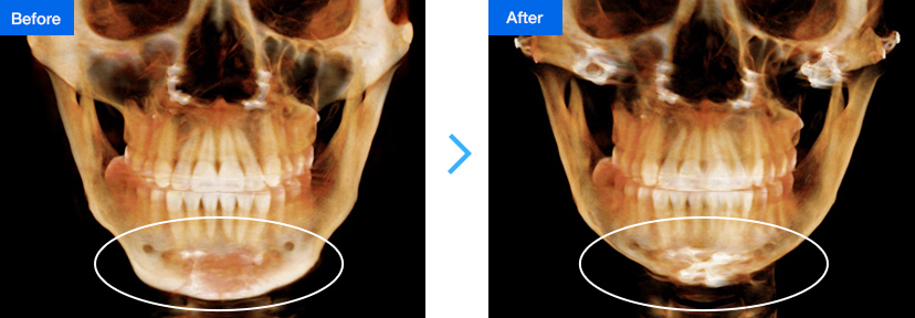
Cases Requiring Revision Genioplasty
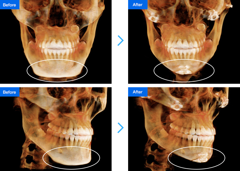
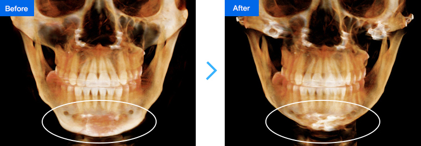
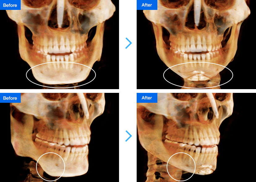
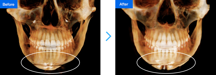
1. No reduction seen from the front after square jaw surgery
case 1
The following are 3D-CT images of real patients who
had revision surgery from Chief Dr. Kim Tae-Gyu.
After primary square jaw surgery using jaw-shaving technique, the chin was still wide and long; V-line square
jaw surgery using inverted V-shape osteotomy and lateral cortical osteotomy were performed for revision.
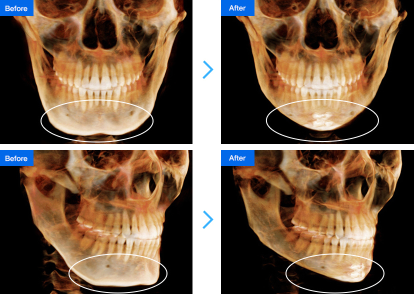
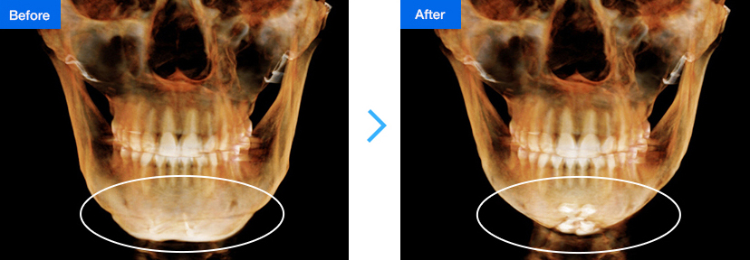
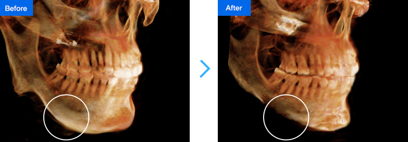
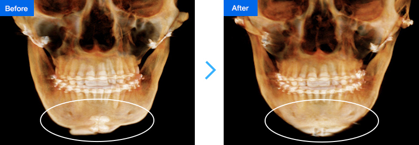
case 2
The following are 3D-CT images of real patients who
had revision surgery from Chief Dr. Kim Tae-Gyu.
After primary square jaw surgery using jaw-shaving technique, no reduction is seen from the front; V-line
square jaw surgery using inverted V-shape osteotomy and lateral cortical osteotomy were performed for revision.
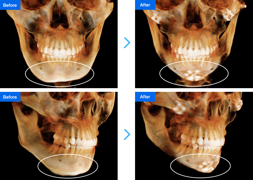
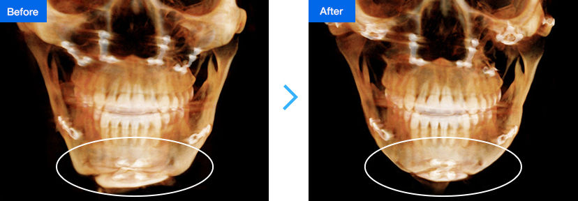
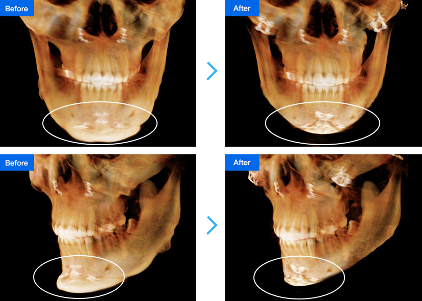
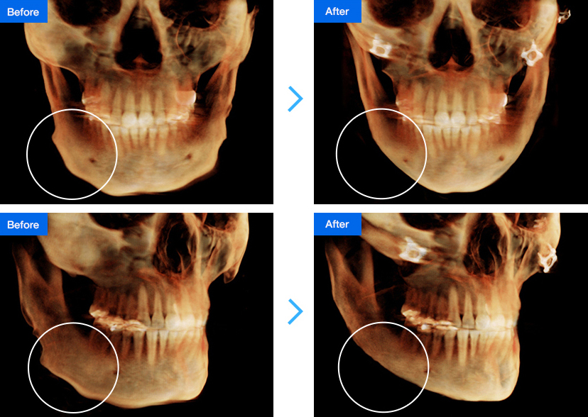
case 3
The following are 3D-CT images of real patients who
had revision surgery from Chief Dr. Kim Tae-Gyu.
After primary square jaw surgery using jaw-shaving technique, no reduction is seen from the front; V-line
square jaw surgery using inverted V-shape osteotomy and lateral cortical osteotomy were performed for revision.
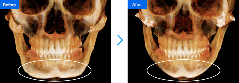
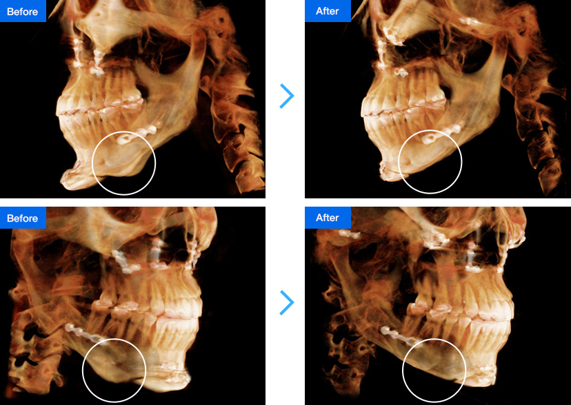
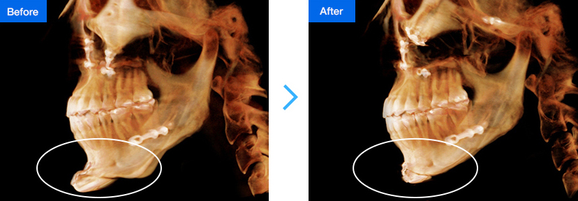
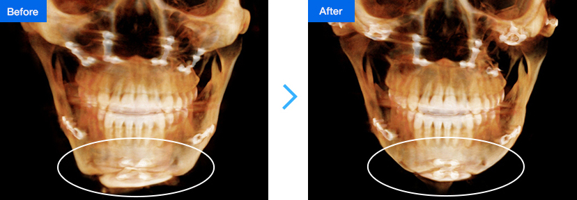
case 4
The following are 3D-CT images of real patients who
had revision surgery from Chief Dr. Kim Tae-Gyu.
After primary square jaw surgery using jaw-shaving technique, no reduction is seen from the front and
asymmetry remains; anterior square jaw surgery and lateral cortical osteotomy were performed for revision.
2. Seeking a slimmer face even after V-line surgery (T-shape osteotomy)
case 1
The following are 3D-CT images of real patients who
had revision surgery from Chief Dr. Kim Tae-Gyu.
After primary square jaw surgery using jaw-shaving technique, no reduction is seen from the front and
asymmetry remains; anterior square jaw surgery and lateral cortical osteotomy were performed for revision.
case 2
The following are 3D-CT images of real patients who
had revision surgery from Chief Dr. Kim Tae-Gyu.
Primary V-line square jaw surgery using T-shape osteotomy neither made a reduction seen from the front
nor corrected the wide and long chin; V-line square jaw surgery and lateral cortical osteotomy were
performed for revision.
case 3
The following are 3D-CT images of real patients who
had revision surgery from Chief Dr. Kim Tae-Gyu.
Primary double jaw and V-line surgery using T-shape osteotomy made no reduction seen from the front
and an irregular chin shape remained; V-line square jaw surgery using inverted V-shape osteotomy and
lateral cortical osteotomy were performed for revision.
case 4
The following are 3D-CT images of real patients who
had revision surgery from Chief Dr. Kim Tae-Gyu.
Primary square jaw surgery using T-shape osteotomy neither made a reduction seen from the front nor
corrected the wide chin; V-line square jaw surgery and lateral cortical osteotomy were performed for
revision.
3. Creation of secondary angle or staircase-shaped jaw
case 1
The following are 3D-CT images of real patients who
had revision surgery from Chief Dr. Kim Tae-Gyu.
Primary double jaw and chin surgery made an irregular and staircase-shaped jaw line; V-line square jaw
surgery using inverted V-shape osteotomy and lateral cortical osteotomy were performed for revision.
case 2
The following are 3D-CT images of real patients who
had revision surgery from Chief Dr. Kim Tae-Gyu.
Primary square jaw surgery produced a secondary angle; V-line square jaw surgery using inverted V-shape
osteotomy and lateral cortical osteotomy were performed for revision.
case 3
The following are 3D-CT images of real patients who
had revision surgery from Chief Dr. Kim Tae-Gyu.
Primary square jaw surgery produced a secondary angle; V-line square jaw surgery using inverted V-shape osteotomy and lateral cortical osteotomy were performed for revision.
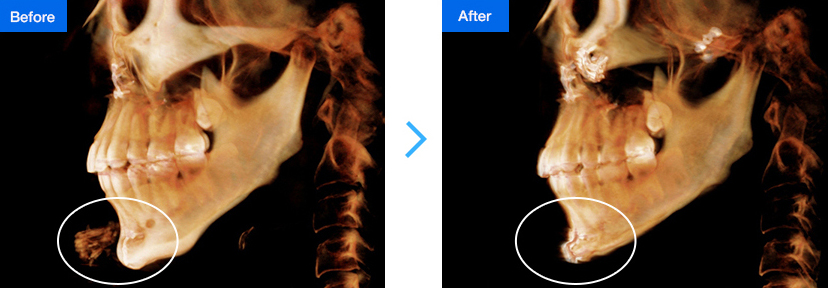
4. Elongated or protruded chin
case 1
The following are 3D-CT images of real patients who
had revision surgery from Chief Dr. Kim Tae-Gyu.
Primary mouth protrusion surgery with T-shape osteotomy was unable to shorten the chin and caused it
to protrude; V-line surgery using inverted V-shape osteotomy and reverse genioplasty were performed for
revision.
case 2
The following are 3D-CT images of real patients who
had revision surgery from Chief Dr. Kim Tae-Gyu.
Primary double jaw surgery and advancement genioplasty made the chin longer and protruded; V-line
surgery using inverted V-shape and reverse genioplasty were performed for revision.
case 3
The following are 3D-CT images of real patients who
had revision surgery from Chief Dr. Kim Tae-Gyu.
Primary T-shape osteotomy was unable to correct the wide and long chin; V-line square jaw surgery using
inverted V-shape osteotomy and lateral cortical osteotomy were performed for revision.
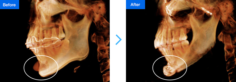
5. Shortened Chin
case 1
The following are 3D-CT images of real patients who
had revision surgery from Chief Dr. Kim Tae-Gyu.
Primary T-shape osteotomy shortened the chin and aggravated its asymmetry; a chin bone graft, anterior
square jaw surgery and lateral cortical osteotomy were performed for revision.
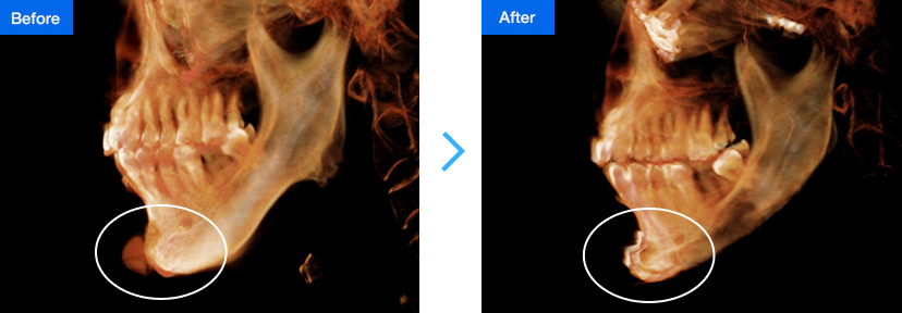
6. Uneven or irregular chin and jaw line
case 1
The following are 3D-CT images of real patients who
had revision surgery from Chief Dr. Kim Tae-Gyu.
Primary square jaw surgery made the chin and jaw line irregular; anterior square jaw surgery and lateral
cortical osteotomy were performed for revision.
7. Unchanged or aggravated asymmetry
case 1
The following are 3D-CT images of real patients who
had revision surgery from Chief Dr. Kim Tae-Gyu.
Primary double jaw and V-line surgery using T-shape osteotomy aggravated the asymmetry; V-line square
jaw surgery using inverted V-shape and lateral cortical osteotomy were performed for revision.
case 2
The following are 3D-CT images of real patients who
had revision surgery from Chief Dr. Kim Tae-Gyu.
Primary V-line surgery using T-shape osteotomy aggravated the asymmetry; chin reconstruction using
artificial bones and osteobond was performed for revision.
8. Seeking bone surgery with implant or filler removal
case 1
The following are 3D-CT images of real patients who
had revision surgery from Chief Dr. Kim Tae-Gyu.
Primary filler injection made the chin reddened and too long; V-line surgery using inverted V-shape
osteotomy and advancement genioplasty were performed for revision.
case 2
The following are 3D-CT images of real patients who
had revision surgery from Chief Dr. Kim Tae-Gyu.
Primary chin implant surgery made the chin thicker and unnatural; V-line surgery using inverted V-shape
osteotomy and advancement genioplasty were performed for revision.
case 3
The following are 3D-CT images of real patients who
had revision surgery from Chief Dr. Kim Tae-Gyu.
Primary chin implant and square jaw surgery made the chin too long and protruded; V-line surgery
using inverted V-shape osteotomy and advancement genioplasty were performed for revision.


![2013年消费者最值得信赖的品牌大奖
[鼻部整形/面部轮廓整形领域]荣获大奖](/images2/main/partners_000.jpg)
![2013年韩国消费者最受欢迎品牌第一名
[鼻部整形/面部轮廓整形领域]荣获大奖](/images2/main/partners_03.jpg)